

Equine Foetal Sexing
Veterinary Ultrasound Imaging diagnosis and monitoring scans
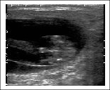
What is veterinary ultrasound maging? Technique and basic principles of ultrasound. Veterinary monitoring & diagnostics Imaging. Technology advantages of routine investigative scans, ultrasound guidance.