Advantages of use and undertanding Veterinary Ultrasound by machine modes, settings, applications, applicators e.g. transducers, types and features of transducers sutability of probes and scanning machines. practical clinic and field based ultrasound examinations.

Archive and reporting
Comprehensive Guide on Veterinary Imaging Examinations: Documentation, Compliance, and Enhanced Diagnostics Veterinary Examinations: Veterinary imaging examinations play a crucial role in modern veterinary medicine, offering invaluable insights that aid in…
presets of ultrasound scanner machines
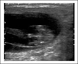
B Mode Ultrasound basics of veterinary ultrasound scanning

Equine Foetal Sexing
What is veterinary ultrasound maging? Technique and basic principles of ultrasound. Veterinary monitoring & diagnostics Imaging. Technology advantages of routine investigative scans, ultrasound guidance.