
Mare Pregnancy


Pregnancy in the Mare:
Embryo Implantation:
Our scanners for examinations of pregnancy in the Mare:
MOBILITY phase in uterine horns and body until Day 16: FIXATION (termination of mobility) then occurs. Fixation should be at the base of one of the uterine horns. This early mobility of the equine conceptus is important to block endometrial prostaglandin release. The prostaglandin arrives via a systemic pathway as the mare does not have a close connection of the uterine vein and ovarian artery
MPLANTATION begins around Day 40 and is not complete until around Day 140 when formation of the micro-cotyledons is complete. Attachment of the placenta is gradual in the mare and is unable to be precisely temporally defined
ENDOMETRIAL CUPS: Distinctive, irregular-shaped areas which develop to become grossly visible arranged in a ring at the base of the pregnant horn. Begin to produce equine chorionic gonadotrophin (eCG; old term PMSG) and appears in the circulation around Day 35. Values rise rapidly to peak around 65 Days. There is then a variable rate of decline, but may persist as long as 150 days. This effectively means the mare cannot be bred again that year once she is positive for eCG. eCG is responsible for ovulation and/or luteinisation of secondary ovulations.
Progesterone Sources: Until Day 200: primary and secondary corpora lutea
From Day 60 : Feto-Placental Unit produces progestins and gradually assumes the main role
Equine Uterine changes: becomes tonic (17 -21 Days) and swelling develops about 21 – 24 Days; by 30 Days the embryonic vesicle is 3 to 4 cm in diameter and by 40 Days is the size of a tennis ball; between Days 60 and 100, the uterus is low within the abdomen and the foetus can not usually be palpated; from 4 to 5 months onwards the foetus can usually be palpated
Equine Cervix changes:Tight, firm and tonic (as during dioestrus)
Ovarian Follicle: Ovarian changes: 18 to 40 Days – many follicles up to 3 Cm, occasional ovulations; 40 to 120 Days – extensive ovarian activity with ovulations; luteinisation and development of secondary corpora lutea; 120 days to term: small and inactive, difficult to palpate after 5 months
Duration of Equine pregnancy is 330 to 345 Days, but enormous variation is possible and anywhere from 315 to 360 Days is frequently reported
Diagnosis of early pregnancy using ultrasound: Day 11
• The equine embryonic vesicle can be reliably detected at day 11 when sufficient anechoic yolk sac fluid has developed.
• The black yolk sac fluid is enclosed within the trophoblast resulting in a spherical outline with a diameter of 5 to 8 mm.
• Note the two hyperechoic short lines on the dorsal and ventral borders of the conceptus known as specular reflections. Whilst they may aid location of the early embryonic vesicle, they are not indicative of pregnancy but are a physical phenomenon arising from the reflection of the ultrasound beam; cysts tend to cause non-specular reflections
• The anechoic vesicle is highly mobile at this stage and found in either horn or in the body. It is, therefore, important to examine both uterine horns and the body thoroughly.
Mares are not usually scanned as early as Day 11 because it is possible to miss the conceptus if scanning conditions are not ideal and the ovulation date is not accurately known. If there was an ovulation one or two days after the first ovulation, any pregnancy arising from this later ovulation would be too small to be detected.
Diagnosis of early pregnancy using ultrasound: Day 14
• The 14 day conceptus is 13 to 18 mm in size and lies centrally in the uterine body.
• Note the spherical shape and increase in size over the Day 11 pregnancy. The embryonic vesicle grows at a rate of approximately 3.5 mm/day at this early stage of pregnancy and remains highly mobile, making thorough examination of all parts of the uterus important.
• In the event of twin pregnancies, both vesicles can usually be seen at 14 days, even if the second co-twin arose from a later ovulation.
• This fact, together with the mobility and relatively small size of the concepti make 14 to 15 days the optimal stage of pregnancy to diagnose twins and crush one co-twin.
Although pregnancy diagnosis is highly accurate even at this early stage, it is important to be aware of the possible confusion caused by uterine cysts and the presence of twin conceptuses. Ideally one would have performed an ultrasound examination before breeding the mare, but this is not always possible. If the first scan is performed at Day 14 or 15, then it is possible to return the next day in cases of confusion and see if the pregnancy has changed position or grown in size. This should allow differentiation from a cyst before the pregnancies have a chance to become unilaterally fixed.
Another advantage of performing the first examination for pregnancy at Day 15 and not Day 18 is that a mare with a shortened luteal phase due to endometritis can be detected. If examination is delayed until Day 18 it is possible that the mare has ovulated already and the fact that she has had a shortened luteal phase will not be detected.
Many veterinarians point out to me that it may be better to delay the first examination until Day 18 when non-pregnant mares have had the opportunity to return to oestrus and it may be possible to predict the next breeding time. However it will be very difficult to correct unilateral twins. Whilst there is undoubtedly a natural reduction mechanism of twin pregnancies to singletons in mares, I believe that the effectiveness of this has been exaggerated and is not as successful as manual crushing of one twin at Day 15 which has a 95% success rate in my experience.
Double ovulations occur during 8 to 30% of oestrous cycles, the frequency depending upon the breed and type of the mare (Thoroughbreds, highest rate; ponies, lowest rate). Accurate detection of such ovulations is important as twinning is undesirable, firstly because it accounts for 10–30% of abortions and, secondly, even if both foetuses survive and are carried to term, many are dysmature, resulting in a high neonatal mortality rate. A further complication is that if embryonic/foetal death occurs after the formation of the endometrial cups these latter structures persist until they spontaneously regress as if pregnancy had been maintained, resulting in pseudo-pregnancy.
Effective twin management is a vital part of maximising fertility on the stud farm. In my experience owners have accepted the early pregnancy check when the reasoning has been explained to them. Of course, if the mare must be transported back to the stud farm or to the veterinarian for scanning this may present more of a difficulty.
It is important to realise that if twin pregnancies arise from asynchronous ovulations, once conceptus is likely to be larger than the other. This is not an indication to leave the smaller pregnancy, manual crushing should still be performed
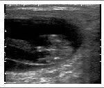
By day 16 of equine pregnancy
the vesicle is normally fixed at the base of either the left or the right horn. The shape is still regular, but more ovoid than strictly spherical.
At day 20 of pregnancy the conceptus is irregular in outline. This irregular shape is normal and is not an indication of imminent pregnancy loss. The appearance can be so irregular that it could be confused with a collection of intraluminal uterine fluid, particularly as no embryo is likely to be visible at this stage. Therefore, a single pregnancy scan at Day 20 is a bad idea. Note the thickened dorsal wall of the uterus and the presence of a slight oedema pattern above the conceptus. This may be due to oestrogen production by the conceptus or the follicular development typical of this stage of pregnancy. If the pregnancy is normal, the primary corpus luteum should also be visible. If the oedema pattern of the uterus becomes widespread it may be an indicator of early embryonic death (see later). It is important to re-schedule subsequent pregnancy examinations at more frequent intervals than normal and the first re-examination should be in 3 or 4 days to monitor the pregnancy.
By day 24 of equine pregnancy, the embryo is approximately 6 mm in length. The heartbeat can normally be detected as a flickering movement in the middle of the echoic embryo around this stage of pregnancy. Note the emergence of the allantoic sac as a small anechoic area from beneath the embryo. Over the next few days, the development of the allantois will lift the embryo dorsally and the yolk sac will gradually reduce in size. This change in ratio of the yolk sac to allantois is an important feature in ageing pregnancies. It is important to recognise the embryo and identify a heart-beat because the irregular shape of the vesicle is easily confused with an endometrial cyst.
At day 20 of equine pregnancy the conceptus is irregular in outline. This irregular shape is normal and is not an indication of imminent pregnancy loss. The appearance can be so irregular that it could be confused with a collection of intraluminal uterine fluid, particularly as no embryo is likely to be visible at this stage. Therefore, a single pregnancy scan at Day 20 is a bad idea. Note the thickened dorsal wall of the uterus and the presence of a slight oedema pattern above the conceptus. This may be due to oestrogen production by the conceptus or the follicular development typical of this stage of pregnancy. If the pregnancy is normal, the primary corpus luteum should also be visible. If the oedema pattern of the uterus becomes widespread it may be an indicator of early embryonic death (see later). It is important to re-schedule subsequent pregnancy examinations at more frequent intervals than normal and the first re-examination should be in 3 or 4 days to monitor the pregnancy.
In the day 28 pregnancy
note the developing allantois, the regressing yolk sac and the associated dorsal ‘ascent’ of the embryo. The apposition of yolk sac and allantois results in an ultrasonically visible thin line normally orientated horizontally. The embryo is visible as an echoic mass on this line.
By day 30 of pregnancy note the enlargement of the allantoic sac such that the two sacs are approximately equal in size. The embryo is highly echogenic and is visible on the line separating the allantoic and yolk sacs and the heartbeat can be clearly seen.
day_30jpg goes here!
By day 35 the embryo is usually in the dorsal part of the vesicle and is approximately 16 mm in length. The volume of the allantois greatly exceeds that of the yolk sac.
day37.jpg goes here!
Pregnancy Diagnosis: Protocol (When Ovulation Time is Known)
First exam Day 14 to 15
Second exam Day 24 to 27
Third exam Day 33 to 35
Autumn exam October
Veterinary applications Equine Equine Pregnancy
(day of ovulation is Day 0)
Following an initial examination at Day 15, the aim of the examination at Day 24 to 27 should be to assess the embryo is developing normally (increase in size, normal echogenicity of the yolk sac and so on) and identify the heartbeat. In addition it can be conformed that there is only a single conceptus. If twins were inadvertently missed at the earlier examination, it may still be possible to correctly manage them.
Ideally a third examination should be performed around Day 33 to 35. The aim of this examination is to confirm that a single conceptus is developing normally. If there is failure of normal development or if twins are detected, it is usually possible to terminate the pregnancy and re-breed the mare.
If examination is delayed until after day 33, the endometrial cups may have developed and even if pregnancy is terminated, eCG production may continue for a variable time, sometimes preventing normal oestrous cycles for the rest of the breeding season. In addition much time may be lost if early embryonic death has occurred and earlier scanning will allow earlier detection of this loss.
These timings are only suggestions and a routine must be established which is applicable to your particular situation and also the mare involved. Some mares, for example mares with several uterine cysts, may require additional examinations.
NB:
1. From older studies and lactures of Mr Pycock with Pie data, when images were taken using the Scanner 100 LC Vet built-in floppy disk drive)
2. New scanners have more features and image detailed defined images then represented here
Ultrasound scanner for Pregnancy in the Mare: from Pie Data UK Ltd
Our scanners for examinations of pregnancy in the Mare: