What is Veterinary Ultrasound Imaging? Diagnosis and monitoring scans
What is Veterinary Ultrasound Imaging? Diagnosis or monitoring scans

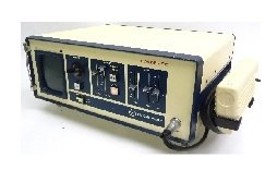
Use of Ultrasound scanners in veterinary medicine is widely used since early 1970’s, following Sonar technology on ships and fishing where soft tissue images are created by using sound waves that are emitted from a transducer and then bounce off. Distance is detected. similarly in animal and human medicine, soft tissue, fluid and bones depending on its density sound waves are reflected from the animal’s area of examination.

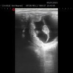
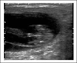
These frequency dependent sound waves are used to create different grey values to build the ultrasound data for the reflected image, which are then converted into a real-time anatomical image that can be used for diagnostic purposes.

The scanned image produced by ultrasound is incredibly useful for veterinarians, as it allows them to visualize both normal and abnormal structures within the animal’s body, aiding in the diagnosis of various conditions and diseases. As an example for equine reproduction stages of pregnancy can be monitored for mare’s and fetal health during fertility to birth of a foal.

Various modes can be changed on a well designed portable ultrasound machine that can provide various types of none invasive clinical examinations as by examinations of anatomical structures are to be performed using ultrasound technology.

